- Rhinoplasty
- Sinusitis Treatment
- Revision Rhinoplasty
- Bone Curvature (Septum Deviation)
- Septum Perforation Treatment
- Nasal Flesh (Concha Hypertrophy)
- Nasal Congestion
- Hearing Loss
- Cochlear Implant
- Hearing Loss in Children
- Tympanoplasty
- Vertigo
- Tinnitus
- Middle Ear Microsurgery
- Otosclerosis
- Ear Tube
- Larynx Cancer
- Trachea Diseases, Stenosis
- Oral Cancer
- Cancer of the Pharynx
- Salivary Gland Diseases, Tumours
- Thyroid Surgery
- Neck Masses
Contents
ToggleOral Cancers: Diagnosis, Treatment and Symptoms
The mouth is an anatomical structure consisting of two cavities: vestibulum and cavum oris. The vestibulum oris refers to the groove-shaped space between the inner surface of the lips and cheeks and the outer surfaces of the teeth, while the cavum oris describes a large area surrounded by the teeth in front, the tonsillar ring behind, the palate above and the floor of the mouth below. The oral cavity consists of different anatomical regions such as the lips, cheek mucosa, retromolar trigone, upper and lower alveolar arch, floor of the mouth, hard palate and tongue.
Cancer represents a malignant tumour that develops from cells in the layers that cover the body from the outside and inside, called epithelium. Cancerous tumours grow by destroying surrounding tissues and can metastasise to the lymph nodes and other organs of the body. Benign tumours, on the other hand, usually grow without damaging the surrounding tissues and do not spread to the lymph nodes or other organs.
Oral cancers can be seen in the following areas:
- Mouth base
- Language
- Gum and jaw bone
- Cheek (buccal mucosa)
- Tonsil (tonsil)
- Hard or soft palate
- Lips
Risk factors for oral cancers can be the following:
- Smoking and use of tobacco products
- Excessive alcohol consumption
- Tooth decay
- Improper placement of prostheses
- HPV (Human Papilloma Virus) infection, especially types 16-18
– Kötü ağız hijyeni
– Tütsülenmiş gıdaların tüketimi
Symptoms of oral cancers may include:
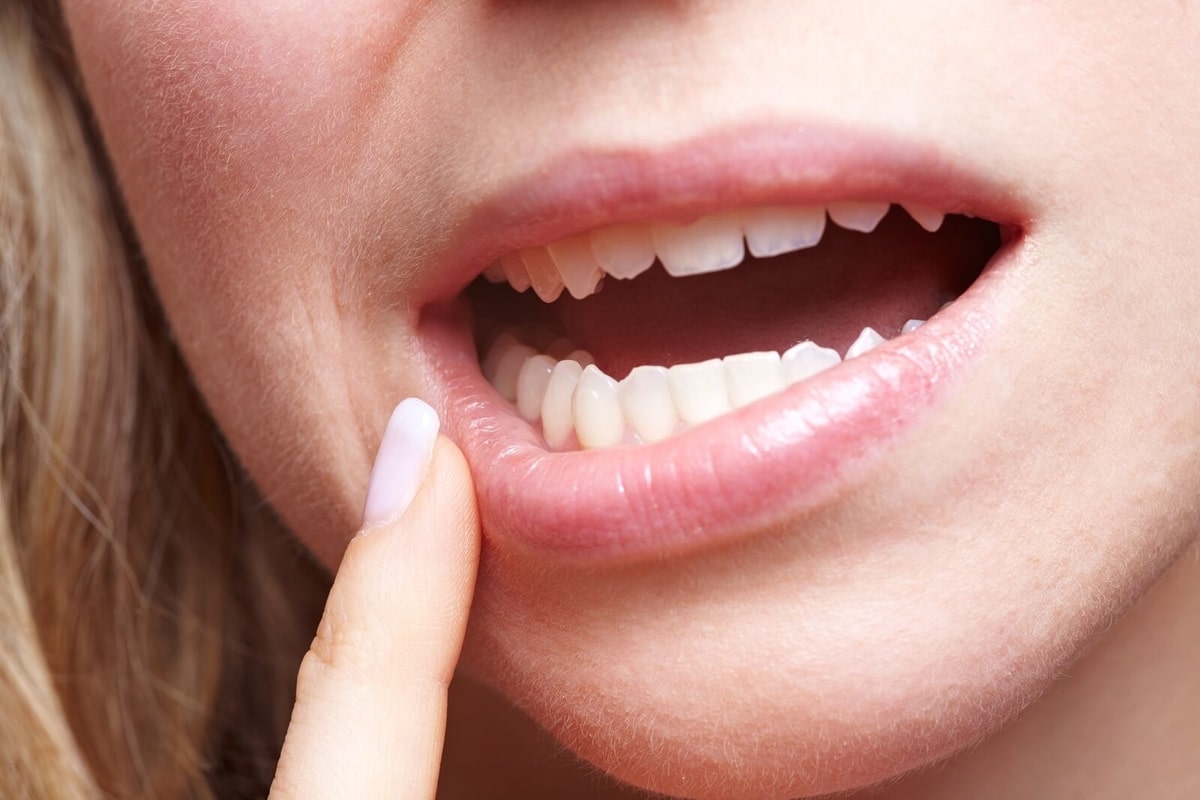
- Wounds on the lips that do not heal for a long time (lasting more than 3 weeks) Color change (red or white spots) in the mucosal tissue covering the inside, swelling or collapse of the mucosa
– Painful or painless swellings in the mouth
– Swelling (mass) in the lymph nodes in the neck
– Speech problems
– Chewing problems
– Painful swallowing (dysphagia)
– Earache
– Loosing or falling out of teeth
– Weight loss
Tongue and intraoral tumours are diagnosed by the following methods:
– Patient's history (when the wound appeared)
– Ear Nose and Throat examination (the depth, width of the wound and the condition of the neck lymph nodes are examined)
– Ultrasound
– Computed tomography (CT)
– Magnetic resonance imaging (MRI)
– Positron emission tomography (PET)
– Examination of scar tissue by biopsy
The treatment of tongue and oral tumours is surgical. Depending on the location, depth and degree of spread of the tumour, resection of the tumour is performed and then the closure of the tumour removal area is planned. Depending on the risk of tumour spread to the neck lymph nodes, neck dissection may also be required. Radiotherapy may be applied depending on the postoperative pathology results. If there is distant metastasis, chemotherapy may be used.
Surgical planning may include:
A) Removal of the tumour, primary closure of the tumour site, neck dissection and postoperative radiotherapy if necessary.
B) Removal of the tumour, neck dissection and closure of the tumour site with a flap, followed by postoperative radiotherapy.
C) Removal of the tumour, neck dissection and closure of the tumour site with a remote tissue transplant (free flap), and postoperative radiotherapy.
In case of distant metastasis, chemotherapy treatment may also be considered.